Background: People Want To Know More Details About Their Intraoral
Nowadays, people are getting younger and younger to developing dental diseases. The dental condition of many young people begins to gradually deteriorate. The reason is that they do not know how to care for their teeth properly. They often ignore the warning signs that tell them that something is wrong with their teeth, even if they have been experiencing some pain or discomfort in their mouths.
When they get older, if they do not take care of their teeth now, then they may need serious treatment later on in life. This will cost them more money than it would have if they had taken care of the problem earlier on in life.
It is better for people to take care of their oral health early on so that they do not have any problems dealing with this situation later on in life when it becomes more expensive and complicated to deal with.
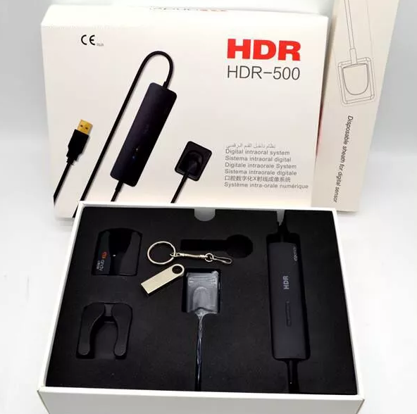
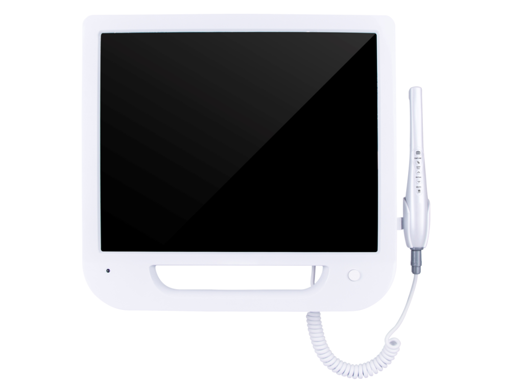
Therefore, there are more and more intraoral cameras that can show a clearer view of the interior of the oral cavity, such as Roson’s intraoral camera.
What Is The Intraoral Camera?
An intraoral camera is a small, hand-held device that produces images of the teeth and gums. It can be used to evaluate the mouth for dental problems and to guide treatment.
The images produced by an intraoral camera are called intraoral radiographs (or “intraos” for short). It is used by dentists and hygienists to examine the teeth and surrounding structures, including the gums, soft tissues, and jaw joints.
What are the 3 types of intraoral radiographs?
Intraoral periapical radiographs:
This type of X-ray shows the teeth and surrounding bone. It is taken from the side or from above, so you may need to move your head or open your mouth wider for this type of shot.
Intraoral bitewing radiographs:
These X-rays show the front surfaces of the upper and lower teeth. They are taken from both sides of your mouth at once, so you do not have to open your mouth too wide for these shots.
Intraoral occlusal (bite) films:
These X-rays show all sides of each tooth at once (front and biting surfaces). They are taken with your mouth closed as well as open, so you may have to move your head around while they are being taken to make sure all surfaces are visible in each photo.
What Are The Four Ways To View An Intra Oral Image?
Roson’s intraoral camera is a camera with a 5-megapixel camera. Its image sensor is CMOS1/4. Its focus range is 5mm–50mm. You can capture details through a specially constructed camera lens (viewing probe).
After it is inserted into the mouth, its light source will make the patient’s oral situation clearer. The picture is then imaged on the image sensor. Finally, you can see a clear enlarged image on the electronic screen. There are four ways to view an intraoral image. They are:
Frontal View:
The frontal view is the most popular because it gives you the best idea of what your teeth look like. It also shows any decay or discoloration that may be present on your teeth.
Occlusal View:
The occlusal view shows the back molars concerning each other, as well as their relationship to the rest of your mouth. This is important because it can reveal problems with your bite, such as over-clenching and grinding.
Lingual View:
The lingual view shows all of the buccal surfaces and incisal edges of your teeth (bordering surfaces). It also shows any decay or discoloration that may be present on these surfaces.
Distal Tip View:
This view is one of two views that allow you to see how long your roots are, which helps determine if there is enough room for an implant or other dental procedure like a crown or bridge placement without fracturing or damaging adjacent teeth or gums during placement of these procedures.
What Are The Common Errors In Intraoral Radiography?
There are many common errors in intraoral radiography. The most common ones are as follows:
- Not positioning the patient correctly. This can lead to distortion of the image and make it difficult to interpret.
- Not focusing on the object at hand (the tooth). The dentist should focus on the tooth and not on the screen, as it is easier to focus on an object that is not moving than on one that is.
- Incorrect exposure setting. If you set it too high, then you will see all kinds of shadows and dark areas in your image; if you set it too low, then there will be no detail or contrast in your image.
Therefore, when observing the patient’s oral condition, it is best to avoid the above mistakes. Only in this way can a clearer and more accurate image be obtained.
Advantages Of Using Roson Intraoral Camera:
The Roson intraoral camera is a high-definition digital camera that is used for diagnosing and treating dental problems. This camera allows you to see inside the mouth with ease and comfort. The advantage of using this device is that it provides clear images of the teeth and gums, which can help in making accurate diagnoses.
Observe the intraoral area clearly:
The main advantage of using Roson’s intraoral camera is that it allows you to observe the intraoral area in real-time. The first advantage is that it can be used for dental treatment, which means that you can observe and record the condition of patients’ teeth during treatment.
Easy to archive and easy to consult:
The second advantage is that it is easy to archive and easy to consult. You can also opt for a camera with WiFi transmission. It will automatically back up to a USB device or send pictures to the website of your clinic or dental office. This way, you will never miss any important details in your patient’s mouth.
Multiple control methods:
The third advantage is that there are many operating systems available with multiple control methods. You can choose between remote control, button control, and mouse control according to your preferences.


